A refined aesthetic sense, creativity, persistence, and a desire to restore a damaged tooth to a healthy and beautiful appearance.
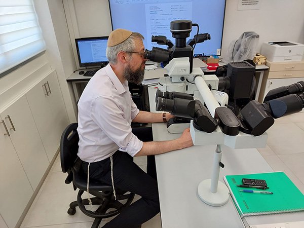
High concentration and use of advanced professional equipment improve visibility for optimal and precise treatment.
Diagnosis and treatment with advanced, pain-reducing methods, alongside professional and compassionate care.
Continuing education and staying up to date with evolving and innovative professional techniques.
As in many fields, current technological advancements enable the development and creation of new and diverse tools that were previously unavailable. In the field of dentistry as well—including procedures related to prosthodontics—such technologies have been integrated to enhance precision and reduce discomfort and unnecessary suffering for patients.
At some point in our lives, most of us (whether personally or through someone close to us) have encountered the process of placing a crown on a damaged tooth. Until a few years ago, this process was long, cumbersome, and unpleasant. It began with the insertion of a measurement tray into the mouth, filled with a silicone-based impression material (similar to putty), to create a model of the tooth—a procedure that caused significant discomfort for many patients. The material was then sent to a dental lab to create the crown model. A considerable amount of time would pass from the moment of the impression to its arrival at the lab, during which the impression material was exposed to temperature changes that could cause dimensional distortion, leading to an imperfect fit of the crown (such as gaps between the crown and tooth, incorrect crown size, etc.).
In recent years, this type of process has become more precise, patient-friendly, and faster. The silicone impression material has been replaced by computers, and the traditional measurement tray by digital scanning. These tools enable accurate three-dimensional imaging of the teeth. The high level of precision is maintained throughout the entire workflow, resulting in a restoration that fits the measured tooth perfectly. The process is relatively simple, non-intrusive, and avoids discomfort for the patient. The time needed to produce a crown has been significantly shortened, as the data is transmitted directly from the scanner to the lab’s computer, and in some cases, the crown can be completed on the same day.
Dental treatment precision is further enhanced through the use of advanced magnification tools. For example, these tools allow the dentist to remove only decayed tooth tissue without affecting healthy tissue or accidentally exposing the dental pulp. Similarly, preparing a tooth for a veneer or crown requires meticulous attention to creating an optimal geometric shape from a mechanical standpoint—goals that can only be achieved with the help of magnification. The modern dentist uses special magnifying glasses (loupes) that enlarge the image of the tooth during the procedure, ensuring conservative and highly accurate work.
In the past, a tooth that had undergone root canal treatment required a post and crown to protect it from fractures. Today, it is known that additional drilling into the tooth root (to place a post) may weaken the root. Moreover, preparing the tooth for a crown removes much of the already limited tooth structure and reduces the long-term survival of the tooth. Placing the crown margin below the gum line increases the likelihood of inflammation and gum recession.
A modern and innovative method uses crowns made of special material (glass-ceramic) known as “endo crowns.” These crowns are chemically bonded to the remaining tooth structure (adhesion), even when it is limited, enabling restoration of the tooth’s function and appearance while preserving as much tooth structure as possible (Endocrowns) and improving prognosis.
This method is also used for natural teeth with extensive crown destruction. In traditional dentistry, treatment of such teeth typically includes root canal therapy, post, and crown. With the new method, a glass-ceramic onlay can be bonded to the existing tooth structure without compromising tooth vitality or sacrificing remaining tooth material.